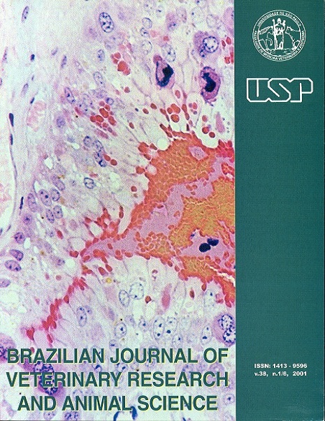
Microscopic features of the umbilical cord in equine (Equus caballus ;¾; Linnaeus, 1758)
DOI:
https://doi.org/10.1590/S1413-95962001000200004Keywords:
Horse, Umbilical cord, Vein, ArteryAbstract
Microscopy of the umbilical cord vein and arteries from cross-bred equine in different pregnancy stages was studied. Umbilical cords from 8 fetuses were collected with age varying from 73 to 249 days. For histological study several staining techniques were used, such as hematoxilin-eosin, Verhoeff, Gordon, picrossirius and Masson's Tricrome. Both the arteries and the veins presented a tunic intern in the constitution of their walls that showed a characteristic plaiting mainly in the arteries, a tunic media that contained a well developed musculature, and a tunic adventitia. Reticular fibers were common characteristics in the wall of the umbilical vessels, even so more numerous in the walls of the umbilical veins. On the other hand, the elastic fibers appeared in small amount mainly in the tunic media and adventitia. Finally, the disposition of the collagen fibers could be evidenced by the picrossirius staining technique, and they were very similar in both the vein and the umbilical arteries.Downloads
Download data is not yet available.
Downloads
Published
2001-01-01
Issue
Section
BASIC SCIENCES
License
The journal content is authorized under the Creative Commons BY-NC-SA license (summary of the license: https://
How to Cite
1.
Carvalho F de SR, Miglino MA, Severino RS, Ferreira FA, Ferreira CG, Santos TC dos. Microscopic features of the umbilical cord in equine (Equus caballus ;¾; Linnaeus, 1758). Braz. J. Vet. Res. Anim. Sci. [Internet]. 2001 Jan. 1 [cited 2024 Apr. 25];38(2):66-8. Available from: https://www.revistas.usp.br/bjvras/article/view/5904


